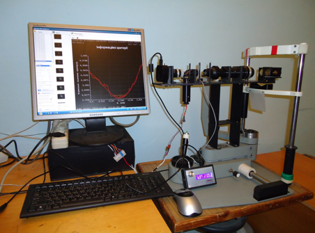
Device for measuring of clinical focus of presbiopic eye
The method of determination of parameters of clinical focus of presbiopic eye is based on the analysis of three-dimensional distribution of luminosity in the zone of geometrical back focus of the optical system of eye. For determination of clinical focus an optical-electronic device, that has the optical system in the complement of that enter, is used: lighting channel, variolens electronically-controlled by her focal distance, Badal system for the optical interface of pupil of eye and variolens, and also video camera for registration of distribution of luminosity in the zone of virtual image of ligt-spot on the retina of eye. A light-spot appears through a lighting channel - laser diode that radiates in a near infra-red range - on a retina in area of fovea. The virtual image of ligt-spot is formed by the optical system of the investigated eye.
Distribution of luminosity in a focal area is determined through the series of videoshots on that luminosity registers oneself in a few ten of sections of virtual image of ligt-spot on a retina. Through the specially worked out algorithm that computer program dependence of geometrical паарметров of focal area, her length, will be determined that volume of pseudoaccommodation.
With the purpose of reduction of sizes of device the analysis of clinical focus can be produced, using the secondary virtual image of clinical focus, that can be created through an additional lens, that is set before the eye of patient on motion rays that go from an eye.
The scan-out of zone of secondary virtual image of clinical focus with the videotape recording of images takes place a speed video camera for a few seconds.

| Attachment | Size |
|---|---|
| 514 KB |


